
- Introduction
- CD2
- CD3
- CD4
- CD5
- CD7
- CD8
T cells belong to the group of white blood cells known as lymphocytes. T Cells have a central role in cell-mediated immunity and are distinguished by their T cell receptor (TCR), a special receptor on their cell surface. T cells originate in the bone marrow, mature in the thymus and travel in the blood to other lymphoid tissues. They are commonly found in areas such as the tonsils, spleen and lymph nodes.
CD or cluster of differentiation cells are present on T cells. Some like CD2, CD3, CD5 and CD7 are pan T cell markers as they are present on most normal mature T cells. Of theT cell markers, CD2 and CD3 are the most specific for T cells. CD5 is strongly associated with T cells but is also expressed on a small subset of normal B lymphocytes and in B-chronic lymphocytic leukemia. CD7 may occasionally be present on early myeloid cells, especially in leukemia. In acute infectious mononucleosis, there is downregulation of the pan T cell markers, namely CD7, and in Sezary syndrome, a T cell cutaneous lymphoma, the T cells express CD4 but do not usually express CD7.
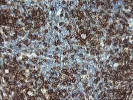
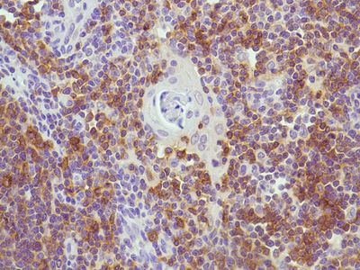
CD 2 is one of the earliest T cell lineage antigens to appear during the differentiation of T cells. It is rare for CD2+ cells to be found in the bone marrow. CD 2 is very useful in immunohistochemistry as it is a Pan T cell antigen marker. CD 2 can also be seen in natural killer cells as well.
CD 2 is very useful for the identification of all normal T cell lymphocytes. This is also useful in the assessment of different types of lymphoid malignancies due to its expression in most precursor and mature T-cell lymphomas and leukaemias. As show below CD2 deletion can also be useful in distinguishing certain tumours, one example being peripheral T-Cell lymphomas. CD 2 can also be used to distinguish T cell from B cell lymphomas. The staining pattern of CD2 is cytoplasmic and membraneous as shown below.
Picture 1 – CD 2 positivity in tonsil
CD 2 has several different applications. Below are a few examples.
Tumour or cell type |
CD2 + or - |
Angioimmunoblastic T cell lympohoma |
Positive |
Lymphoblastic T cell Lymphoma |
Positive |
Cutaneous T cell Lymphoma |
Positive |
Reactive mast cells |
Negative |
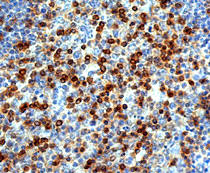
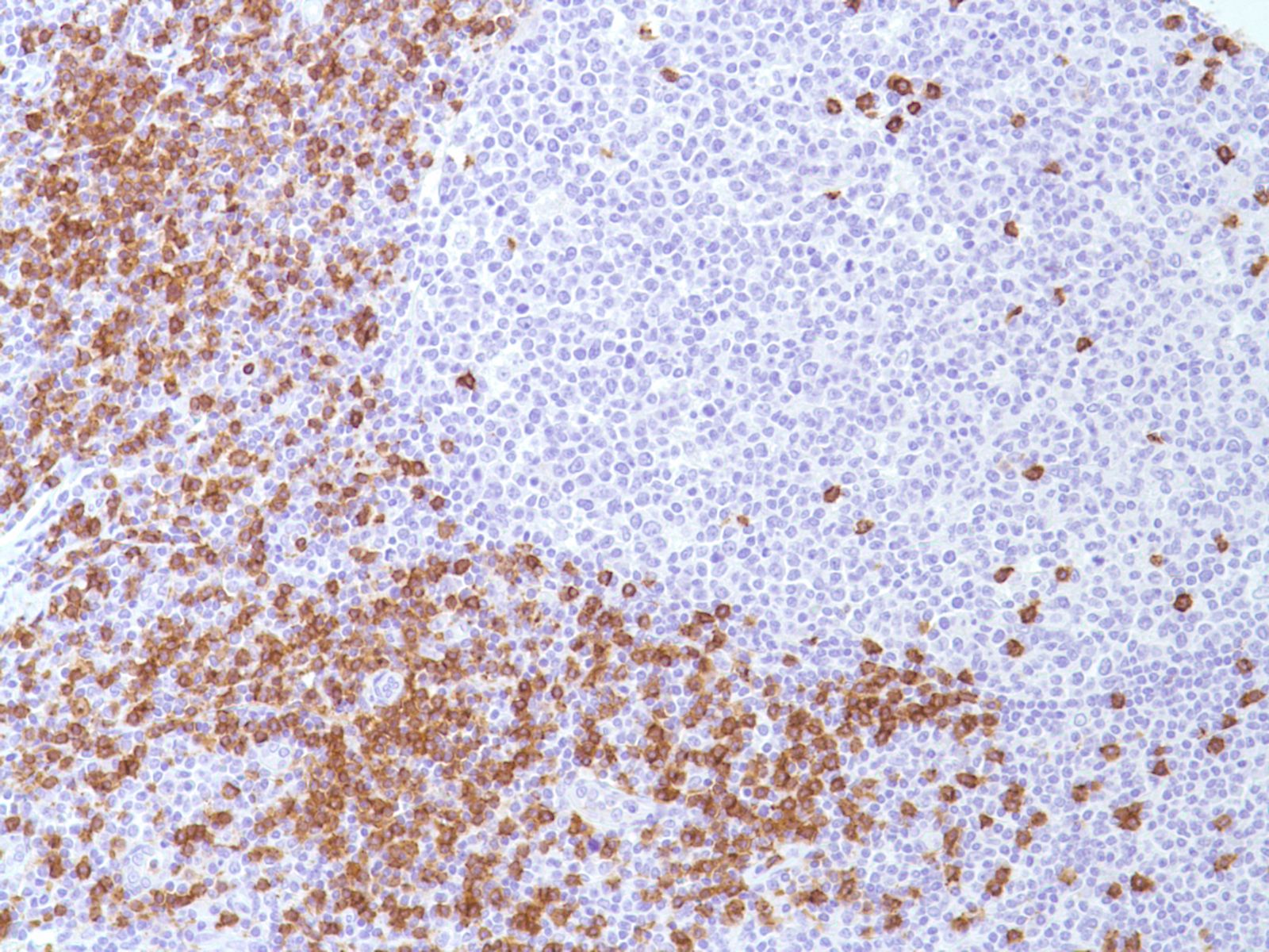
CD 3 is a Pan T cell marker found in the membranes of mature T cells. This is very useful in distinguishing between T cell and B cells origins of tumours. This is also useful in detection of natural killer cells, similar to CD 2. This is one of the most commonly used T cell antibodies in immunohistochemistry as shown in the image below the staining pattern is membraneous.
Picture 2 – CD 3 positivity in tonsil
CD 3 has several different applications. Below are a few examples.
Tumour |
CD 3 + or - |
Mature T cells |
Positive |
Lymphoblastic T cells Lymphomas |
Positive |
Mycosis Fungoides |
Positive |
B Cells |
Negative |
Histiocytic Lesions |
Negative |
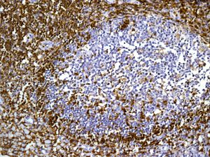
CD 4 is a marker found on T-helper cells, monocytes, macrophages and dendritic cells. CD 4, although not as wide ranging as the Pan T cell marker like CD 2 or 3, is very useful for peripheral T cell lymphomas as the majority of them have origins from T-helper cells and as a result are CD4+/CD8-. As this is a less general T cell marker, CD 4 normally requires other antibodies run in the panel to identify T cell lymphomas
CD 4 is expressed on the surface as well as cytoplasmic as shown in the image below.
Picture 3 – CD 4 positivity in Tonsil
CD 4 has several different applications. Below are a few examples.
Tumour or Cell type |
CD4 + or - |
Angioimmunoblastic |
Positive |
Histocytic lesions |
Positive |
T helper cells |
Positive |
Subcutaneous Panniculitis like T cell lymphoma |
Variable positive in about 85% of cases, but has specificity issues |
Natural Killer cells |
Negative |
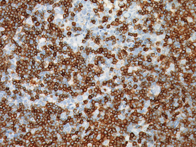
CD 5 is a pan T cell marker, but unlike CD 2 or 3 it also reacts with some B cell tumours like Chronic lymphocytic leukaemia (CLL), Mantle cell lymphoma and also small lyphocytic lymphoma (SLL).
CD 5 can also be used to distinguish between thymic carcinoma and thymoma. As you can see below CD 5 has a membranous staining pattern.
Picture 4 – CD 5 positivity in Mantle cell lymphoma
CD 5 has several different applications. Below are a few examples.
Tumour |
CK AE1/3 + or - |
Hairy cell leukaemia |
Positive |
Mantle cell lymphoma |
Positive |
Thymic carcinoma |
Positive |
T cells |
Variable positive in about 76% of cases, but has specificity issues |
Natural killer cells |
Negative |
Thymoma |
Negative |
CD 7 is pan T cell marker, found in both immature and mature T cells, as well as natural killer cells. This is found in T cell precursors, even earlier than CD2, in myeloid precursors and also in foetal liver and bone marrow. This is particularly useful for lymphoblastic lymphomas and leukaemia as CD 7 is the most commonly expressed T cell marker in these cases.
CD 7 has as shown below a membraneous staining pattern
Picture 5 – CD 7 positivity in Thymus
CD 7 has several different applications. Below are a few examples.
Tumour |
CK AE1/3 + or - |
Angioimmunoblastic T cell lymphoma |
Positive |
Anaplastic T cell lymphoma |
Positive |
Hodgkins Lymphoma |
Positive |
Mycosis fungoides |
Negative |
Adult T cell lymphomas and leukaemia |
Negative |
CD 8 is found on Cytotoxic T cell subset. This is especially useful in combination with CD 4 as it allows differentiation of helper and cytotoxic T cells, allowing a more specific origin of a tumour to be detected.
CD 8 has a membraneous staining pattern as shown below.
Picture 6 – CD 8 positivity in Tonsil
CD 8 has several different applications. Below are a few examples.
Tumour or cell type |
CD 8 + or - |
Cytotoxic T cells |
Positive |
Lymphoblastic T cell Lymphoma |
Positive |
T-cell large granular lymphoma |
Positive |
Anaplastic T cell lymphoma |
Negative |
T-helper cells |
Negative |