Immunohistochemistry- Heavy molecular weight CKs
- Introduction
- Ck 1
- Ck 5
- Ck 10
- Ck 14
- Ck HMW
Cytokeratins are intermediate filaments present in all epithelial cells, and also in a number of non-epithelial cells. In fact, cytokeratin positively has been reported in almost every tumour type, including uterine smooth muscle tumours, most soft tissue sarcomas, melanomas, gliomas, plasmacytomas and occasionally lymphomas.
Cytokeratins can be differentiated into light and heavy chain based on their molecular weight as well as acidic which are cytokeratins 9-20 or basic which are cytokeratins 1-8.
Diagnostic utility
CK 1 is a keratin protein found in humans. This has several uses but as can be seen in Ck5.2, it is most useful when combined with Ck 18.
Picture 1 – CK 1 positivity in Cervix

Ck 1 has several different applications. Below are a few examples.
Tumour |
CK 8 + or - |
Epitheliod sarcoma |
Positive |
GIST or Gastrointestinal Tumour |
Positive |
Angiosarcoma |
Positive in 85% of cases, but has specificity issues |
Karposi Sarcoma |
Variable positive in about 85% of cases, but has specificity issues |
Synovial Sarcoma |
Negative |
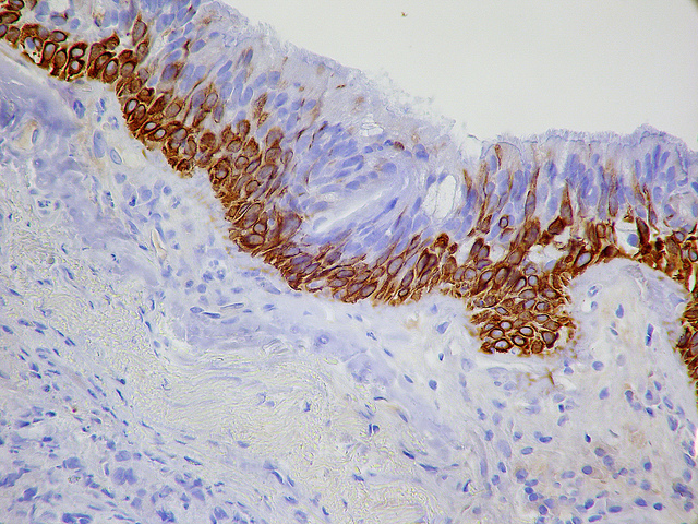
CK 5 is found in most simple ductal and gladular epithelia. A cytoplasmic staining is produced as shown below in the colon but also skin can be used as a control in diagnostic settings. Again this is most useful when combined with CK8.
Picture 2 – CK 5 positivity in bronchial biopsy
CK 5/6 has several different applications. Below are a few examples.
Tumour |
CK 5/6 + or - |
Epitheliod sarcoma |
Positive |
GIST or Gastrointestinal Tumour |
Positive |
Angiosarcoma |
Positive in 85% of cases, but has specificity issues |
Karposi Sarcoma |
Variable positive in about 85% of cases, but has specificity issues |
Synovial Sarcoma |
Negative |
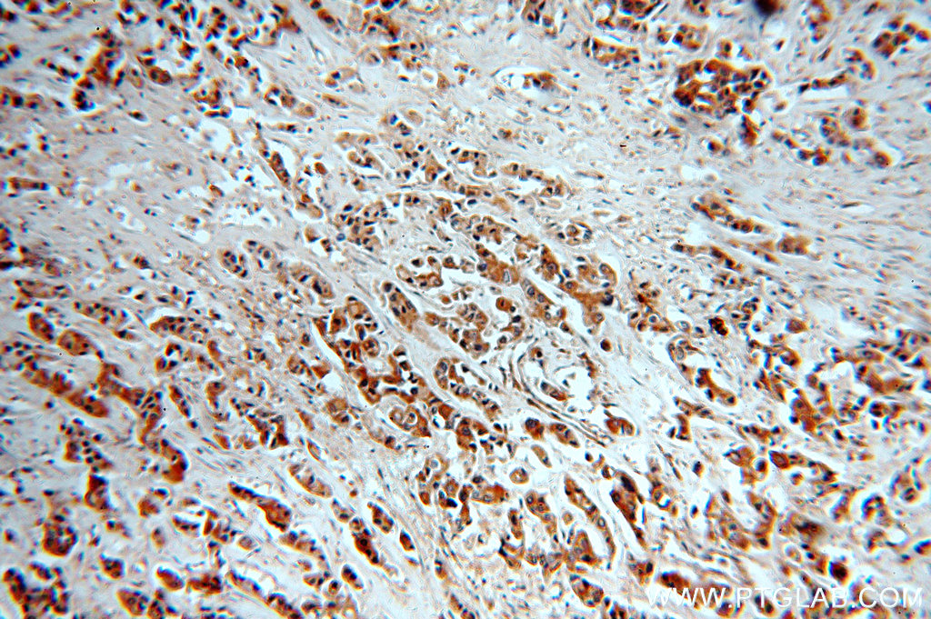
CK 10 reacts with a range of epithelial malignancies. These inclue as shown below adenocarcinomas of colon, stomach, pancreas, liver and breast. A cytoplasmic staining is produced as shown below in the colon but also skin can be used as a control in diagnostic settings.
Picture 3 – CK 10 positivity in breast cancer
CK 10 has several different applications. Below are a few of the most common examples.
Tumour |
CK 19 + or - |
Papillary Thyroid Carcinoma |
Positive |
Thyroid Follicular Carcinoma |
Positive - around 50/60% of the time |
Pancreas Ductal Carcinoma |
Positive |
Neuroendocrine Tumour |
Variable positive in about 50% of cases |
Pancreatoblastoma |
Negative |
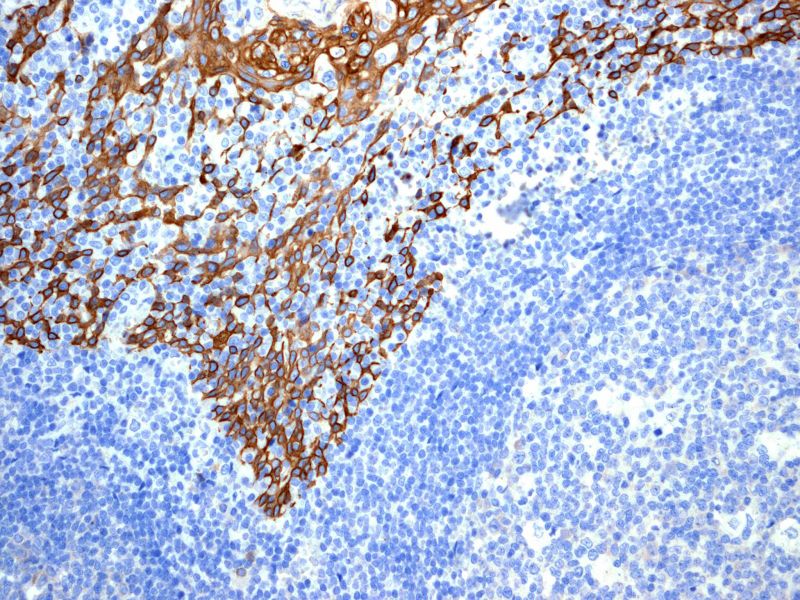
CK 14 has several different applications. It produces a cytoplasmic staining which is very useful in distinguishing adnemocarcinoma and non-keratinizng squamous cell carcinoma from kertainizing squamous cell carcinoma.
Picture 4 – CK 14 positivity in Tonsil
As can be seen below CK 14 has several different applications. Below are a few examples.
Tumour |
CK 14 + or - |
Peripheral nerve sheath |
Positive |
Spindle squamous cell carinoma |
Positive |
Kaposiform Hemangioendothelioma |
Positive |
Keratinizing squamous cell carcinoma |
Negative |
Karposi Sarcoma |
Negative |
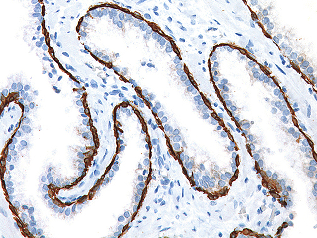
CK HMW or AE3 (Also known as 34BE12) recognises all basic cytokeratins and it is a broadly reactive antibody staining most epithelia and neoplasms. It produces a cytoplasmic staining and is useful in distinguishing Prostatic Adenocarcinoma from Hyperplasia of the Prostate.
Picture 5 – CK HMW positivity in Prostate
As can be seen below CK HMW has several different applications. Below are a few examples.
Tumour |
CK HMW + or - |
Prostatic Hyperplasia |
Positive |
Squamous cell carinoma |
Positive |
Hepatoma |
Positive |
Prostatic adenocarcinoma |
Negative |
Renal Cell carcinoma |
Negative |